Functional and Structural OCT
NIH/NEI R01 EY023285 - PI: Dr. David Huang
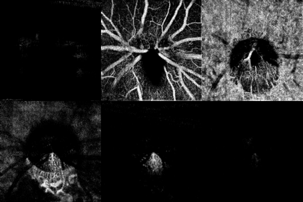
Glaucoma is a leading cause of blindness in the US. The management of glaucoma is based on early detection, followed by careful monitoring to identify those with rapid disease progression and high risk for vision loss. This allows for the rational use of medical, laser, and surgical treatments. Current methods of assessing glaucoma and its progression have significant limitations. Visual field (VF) testing is relatively insensitive during early disease. While quantitative imaging of the optic nerve head and retina with optical coherence tomography (OCT) and other tools are helpful in diagnosis and tracking, their sensitivity and correlation with VF are limited. In the previous grant period, we went beyond conventional thickness analysis of structural OCT and found better ways to analyze nerve fiber layer (NFL) reflectance to improve early glaucoma detection. We have also made fundamental improvements in OCT angiography (OCTA) technology and showed that it can detect and localize glaucoma damage. We developed methods to use OCT and OCTA results to simulate VF to provide objective and accurate measures of glaucoma progression. In the current grant period, we will further improve these promising diagnostic approaches by developing practical means to overcome technological limitations. The Specific Aims are:
1. Develop a directional, high-resolution OCT/OCTA system to improve imaging of structure and perfusion. Our preliminary results have shown that, while analysis of NFL reflectance could greatly improve the detection of early glaucoma, it is limited by variations in the incidence angle of the OCT beam.4 Additionally, OCTA perfusion measurements correlate well with VF parameters and could monitor disease progression, but reliability is limited by variable focusing. We propose to overcome both limitations by developing a directional, high-resolution OCT/OCTA prototype. Real-time control of beam direction ensures the perpendicular incidence necessary for accurate NFL reflectance analysis. Senseless adaptive-optics focusing and aberration correction will be used to achieve reliable high transverse resolution for accurate analysis of nerve fiber bundles and capillaries.13,18 The system will have ultrahigh axial resolution to assess the pentalaminar structure of the inner plexiform layer, a novel approach for glaucoma detection. The prototype will demonstrate that reliable performance can be achieved with real-time system control and optimization using a graphic processing unit (GPU)--an affordable approach that could be used in future commercial clinical OCT systems.
2. Wide-field OCT/OCTA analyses and visual field simulation. Using a fast (120-kHz) commercial OCT system, we will increase the size of peripapillary scans from 4.5x4.5-mm to 6x6-mm, and increase macular scans from 6x6-mm to 9x9-mm. The wider scan will allow visualization of nerve fiber bundle defects from the disc margin to the temporal raphe, which could potentially improve automated detection of early glaucoma defects. Deep learning neural network techniques will be used to combine perfusion, structure, and reflectance maps to optimize the detection of glaucomatous defects. VF simulation will be performed to convert OCTA perfusion measurements and OCT thickness measurements to a VF-equivalent dB-scale. This allows clinicians to assess the speed of glaucoma progression without being misled by the highly nonlinear relationship between raw OCT/OCTA measurements and VF at different disease stages. Peripapillary NFL plexus-based VF simulation provides assessment of broad arcuate sectors that encompass the whole VF. Macular ganglion cell layer plexus-based simulation provides better localization, including the temporal/nasal distinction useful for distinguishing damage patterns of glaucoma from those of other optic neuropathies.
3. Clinical studies in glaucoma diagnosis and monitoring. A longitudinal clinical study will be performed to follow normal subjects as well as open-angle glaucoma patients. These data will be used to validate the above novel OCT/OCTA diagnostic parameters in terms of repeatability and correlation with disease severity as measured by VF and a visual function questionnaire. Both 24-2 and 10-2 VF will be obtained to assess the accuracy of macular VF simulation. This clinical study will test whether combined perfusion, structure, and reflectance analysis can improve the detection of pre-perimetric glaucoma. It will also test whether OCT and OCTA-based VF simulations can better detect glaucoma progression and more accurately measure the speed of progression.
This research is highly likely to transform the clinical practice of glaucoma by developing novel, objective functional and structural tests. OCT is the most widely used ophthalmic imaging test in the US, and OCTA technology has quickly spread since 2014, with implementation on new OCT platforms by at least 4 companies. On newer systems, both OCT and OCTA scans are 3-dimensional and volumetric, making the novel reflectance and perfusion measurements proposed here the logical next step for advancing the use of OCT to evaluate and manage glaucoma in clinical practice.
